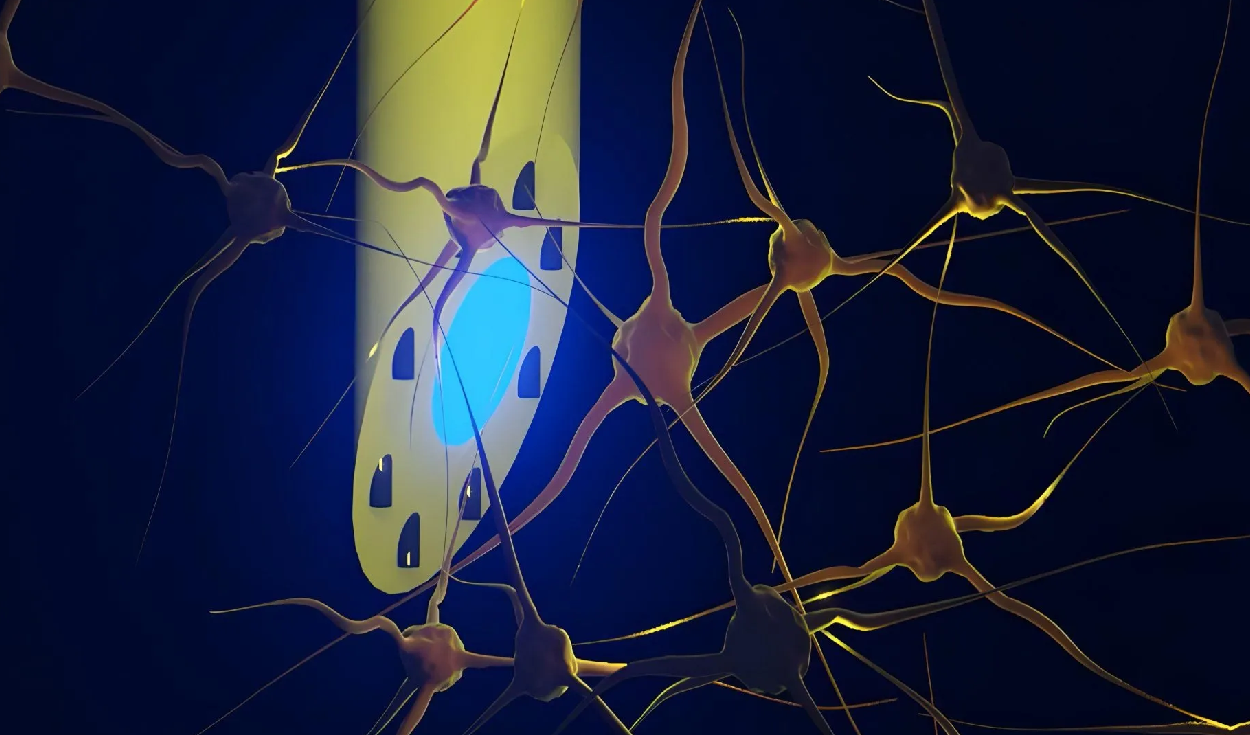
A team of scientists from the Technical University of Denmark, the University of Copenhagen and University College London has developed a flexible, needle-thin brain implant that can perform three distinct functions at the same time. The device, named the microfluidic Axialtrode or mAxialtrode, allows researchers to stimulate nerve cells with light, record electrical signals and deliver targeted substances into specific regions of the brain through a single fiber less than half a millimeter thick.
In initial laboratory tests on live mice, the multifunctional system successfully interacted with both superficial and deep areas of the brain, according to a study published in the scientific journal Advanced Science. The research team designed the ultra-thin fiber to help neuroscientists monitor and manipulate brain activity with unprecedented precision during studies on complex neurological processes.
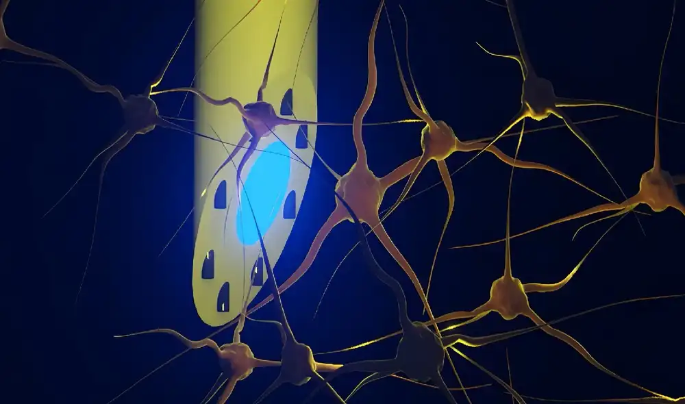
The mAxialtrode measures approximately 420 to 440 micrometers in diameter, making it comparable in width to a fine needle. At its core, the fiber contains an optical channel capable of transmitting light alongside eight microscopic inner channels. These microchannels can transport liquids or house microscopic electrodes designed to pick up electrical impulses from surrounding nerve cells.
Design and Manufacturing of the Fiber
The manufacturing process for the mAxialtrode relies on advanced polymer engineering. Researchers start with a specialized polymer material that is subjected to controlled heat before undergoing a precision stretching process to draw it down to its final microscopic thickness. A micrometer is one millionth of a meter, a scale routinely used in biomedical engineering to fabricate devices that match the tiny dimensions of biological cells and blood vessels.
Unlike conventional optical fibers used in neuroscience research, which typically project light or record signals from a single point at their tip, the mAxialtrode incorporates multiple functional access points distributed along its length. This architecture enables scientists to monitor and stimulate neural tissue across multiple brain depths simultaneously without needing to insert separate probes into the brain.
Traditional brain implants made of rigid silicon or metal often present significant challenges in long-term neurological studies. Because brain tissue is soft and moves slightly with blood flow and respiration, rigid materials can create mechanical friction, causing local tissue irritation and triggering inflammatory immune responses. Developing soft, flexible implants that match the mechanical properties of living tissue has long been a key objective in neural engineering.
Kunyang Sui, a postdoctoral researcher who developed the concept alongside associate professor Christos Markos at the Technical University of Denmark, highlighted the benefits of soft materials in neural probes. Sui explained that rigid implants can irritate brain tissue and trigger inflammatory responses. Sui added that the new device differs because it is constructed from soft, plastic-like optical fibers and features a specially angled tip that reduces overall size and minimizes tissue damage upon insertion into the brain.
Testing in Live Animal Models
To verify the capabilities of the mAxialtrode, researchers conducted in vivo tests on live mice. During the experiments, the single fiber was connected to external light sources, electrical recording instruments and miniature fluidic pumps. The trials confirmed that the device could effectively activate nerve cells using both blue and red light while simultaneously collecting electrical signals across shallow and deep neural structures.
The experiments focused on two crucial brain structures: the cerebral cortex and the hippocampus. The cerebral cortex is the outer layer of neural tissue responsible for high-level cognitive functions, sensory processing and voluntary movement. The hippocampus is a specialized structure located deep within the temporal lobe that plays a central role in memory formation, spatial navigation and learning. Both regions are primary focus areas in studies of brain function and disease.
In addition to optical stimulation and electrical recording, the mAxialtrode demonstrated precise liquid delivery within the brain. Researchers successfully administered different chemical substances to targeted depths in the tissue, using delivery ports separated by up to 2.7 millimeters along the single lightweight strand.
Collaborations and Future Applications
The research brought together specialists from leading academic institutions across Northern Europe. Rune W. Berg led a team at the University of Copenhagen, while Rob C. Wykes headed a group at University College London. The collaborating teams provided essential expertise in neural circuit dynamics and established animal models of epilepsy to test the system in realistic disease environments.
The Technical University of Denmark, located in Lyngby, is widely recognized for its expertise in optical fiber technology and micro-engineering. The University of Copenhagen, founded in 1479, is Denmark's leading medical research institution, while University College London is a world-renowned center for neuroscience research. Epilepsy, one of the primary targets for future applications of the device, is a neurological condition characterized by recurrent, unprovoked seizures caused by abnormal electrical discharges in the brain.
Currently, the mAxialtrode remains in the experimental phase of development. The research team is working to secure patent protection for the novel technology while evaluating the regulatory and technical requirements necessary for potential future clinical trials in humans. Scientists anticipate that the multifunctional fiber could eventually advance research into memory disorders, epilepsy treatments and broader neurological health.