Scientists at the University of Edinburgh have found that life experiences physically reshape the molecular connections between brain cells across more than one hundred regions.
The landmark study, published in the journal Nature Communications, revealed that living in stimulating surroundings does not simply cause the brain to grow more connections. Instead, lived experiences trigger a selective reorganisation of existing pathways, altering the specific identity and anatomical distribution of communication points across the nervous system.
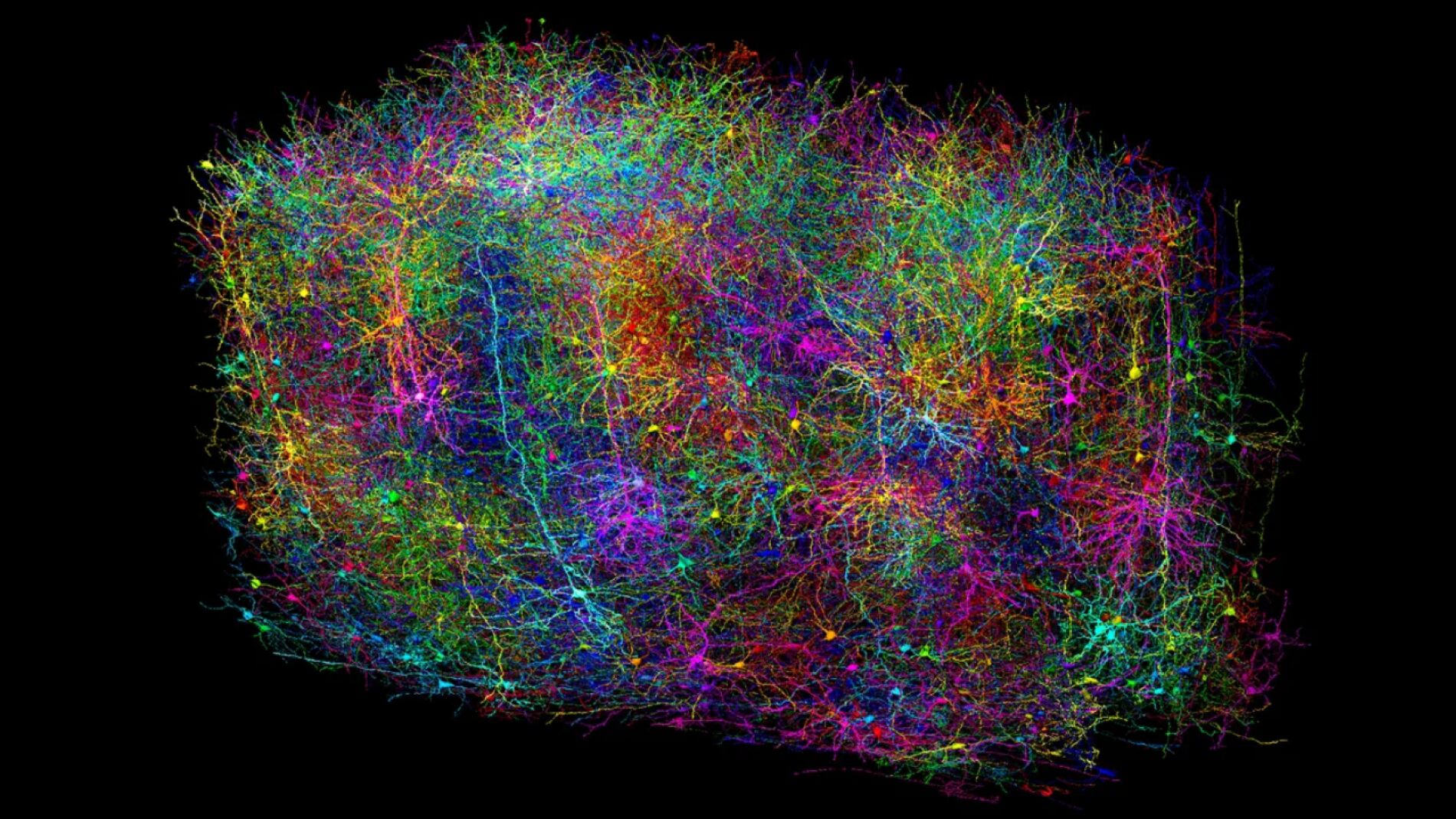
In a healthy human brain, roughly 86 billion neurons form an estimated 100 trillion junction points known as synapses. Neuroscientists often compare this intricate network to a vast metropolitan city, where neurons represent individual buildings and synapses serve as the streets and avenues carrying traffic between them.
Just as city streets vary in size and purpose, different neural pathways handle distinct forms of information. Some carry visual signals from the eyes, while others manage movement, store memories, or process emotional responses. Each brain cell receives incoming messages from numerous neighbours and transmits its own signals through specialised nerve extensions, exchanging information using both electrical pulses and chemical messengers.
Mapping the Synaptome
The overall pattern of these connections, rather than the raw number of neurons, dictates how the brain processes sensory information, learns new skills, and retrieves memories. Mapping this vast communication network is the central goal of connectomics, which remains one of the most significant challenges in modern neuroscience.
To investigate how this network shifts over time, a research team led by Seth Grant used an advanced technique known as synaptome mapping. The synaptome refers to the complete catalogue of different synapse varieties and their precise locations across the brain.
The methodology combines high-resolution microscopy with automated computer image analysis, allowing researchers to identify, catalogue, and examine billions of individual cellular connections. Unlike static anatomical charts, synaptome mapping allows scientists to observe how neural architecture shifts in response to development, learning, injury, and disease.
Selective Reorganisation in Enriched Environments
To test how daily life alters these connections, the Edinburgh researchers exposed laboratory mice to enriched, stimulus-heavy environments. The animals were housed in large enclosures equipped with running wheels, frequently reconfigured mazes, and increased opportunities for social interaction.
The investigators tested two separate groups to evaluate whether age influenced brain malleability. One group lived in the enriched cages during early development, while the second group experienced the enhanced conditions only after reaching full adulthood.
The results demonstrated that environmental stimulation altered specific populations of synapses across more than one hundred distinct brain regions. Crucially, the response was highly varied: certain categories of connections increased in number, other varieties decreased, and some types remained virtually unchanged.
The most revealing finding was that total synapse density did not rise. The brain did not simply construct extra connections to cope with additional activity. Instead, it carefully fine-tuned its molecular structure by choosing which specific synapse types to modify based on the animal's lived experience.
Sensory Inputs and Distributed Imprints
Study co-author Hanan Woods said that sensory and environmental experiences reshape the molecular landscape of synapses throughout the entire brain. She noted that this structural diversity and capacity for adaptation provide an essential framework for understanding how the brain builds resilience and why it may become vulnerable to disease across a lifetime.
The researchers also investigated whether a localised sensory disruption could produce widespread consequences across other areas of the brain. To evaluate this question, the scientists kept one eye closed in a group of mice for seven consecutive days.
The temporary deprivation of vision triggered noticeable alterations in synapse populations. However, the resulting structural shifts were not confined to the visual cortex or regions dedicated to sight. Significant changes also emerged in distant areas responsible for processing other senses and handling memory storage.
The finding proved that a single sensory change leaves a widespread molecular imprint across disparate areas of the brain. The scientists explained that brain functions do not operate within rigid, isolated silos, but work through interconnected networks that continuously collaborate and recalibrate as external circumstances shift.
Screen Use and Future Medical Therapies
Similar patterns of structural adaptation occurred in both developing and mature mice, indicating that the capacity for synaptic reorganisation continues well into adult life.
Professor Grant said that in development and education, the findings raise important questions regarding how diverse combinations of sensory, physical, social, and learning experiences shape neural architecture. He added that the research provides a valuable baseline for future inquiries into how modern environments, social interactions, and prolonged exposure to digital screens on smartphones or tablets alter the brain.
The authors stressed that their findings do not suggest that extended screen use or digital technology is inherently damaging. The study did not conclude that all stimuli produce identical responses, but rather established a new pathway to study how specific activities remodel synapse varieties over time.
Scientists believe that discovering why certain connections adapt while others remain stable will help identify universal mechanisms of brain resilience. These findings could ultimately inform new clinical treatments to repair synaptic networks damaged by physical trauma, environmental hardships, or genetic conditions, underscoring that the brain actively incorporates life experience directly into its physical architecture.